

Posted On: October 26, 2026
Digital X-Rays and Diagnostic Imaging: How Modern Technology Improves Diagnosis Accuracy
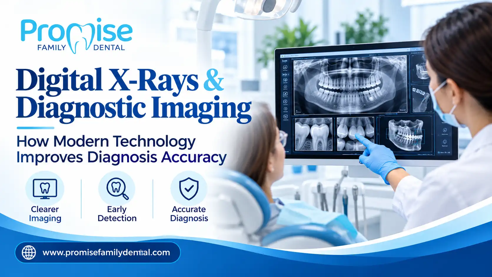
Much of what matters in dentistry happens where you can't see it: between teeth, under old fillings, along the roots, and in the bone that holds everything in place. Digital X-rays and diagnostic imaging let your dentist see those hidden areas clearly and catch problems while they are still small. This guide explains the main types of dental images, how digital imaging compares to traditional film, how often you actually need X-rays, and what to expect at an imaging visit.
What Digital X-Rays and Diagnostic Imaging Reveal That an Exam Alone Can't
A visual exam shows the surface of your teeth and gums. Imaging shows what sits beneath and between them. Dentists rely on it to find:
- Cavities forming between teeth or under existing fillings and crowns
- Bone loss from gum disease
- Infection at the root tip of a tooth
- Impacted or developing teeth, including wisdom teeth
- Cracks, cysts, or other changes in the jaw that don't cause symptoms yet
Common Types of Dental Images and What Each One Is For
Bitewing X-Rays
Bitewings show the upper and lower back teeth together in one view. They are the standard tool for spotting decay between teeth and checking the bone level around them.
Periapical X-Rays
These capture one or two teeth from crown to root tip, along with the surrounding bone. They are commonly used when a tooth is painful or when a root problem is suspected.
Panoramic X-Rays
A panoramic image shows the entire upper and lower jaw in a single sweep. It is useful for evaluating wisdom teeth, planning orthodontic care, and getting a broad view of the jaw.
3D Imaging (Cone-Beam CT)
Where a case calls for it, 3D imaging gives a detailed volume of the teeth, bone, and nerve pathways. It is most often used for implant planning, impacted teeth, and complex root anatomy, and is available at practices equipped for it.
How Digital Imaging Improves Diagnosis
- Immediate results — images appear on screen within seconds, so findings can be reviewed with you during the same visit
- Adjustable views — brightness, contrast, and magnification can be adjusted to examine a suspicious area more closely
- Easy comparison over time — stored images can be compared side by side to see whether a spot is stable or changing
- Simple sharing — images can be sent to a specialist without printing or physical copies
- No film chemicals — there is no chemical developing step or film to dispose of
Digital vs. Traditional Film: Side-by-Side Comparison
How Often Do You Actually Need X-Rays?
There is no single schedule that fits everyone. National guidance asks dentists to base the timing on each patient's risk of decay, gum disease, and other factors, and to adjust it as that risk changes. In practice:
- Patients with active cavities or higher decay risk are imaged more often
- Patients with healthy teeth and low risk can go longer between bitewings
- New patients usually get a baseline set so future changes can be compared against it
- Specific problems, like a toothache or swelling, may call for an image outside the normal schedule
A Dentist in Suffern who explains why a particular image is being recommended is following this approach, and you are always welcome to ask what a given X-ray is meant to find.
What the Data Shows
Dental X-ray timing is meant to follow your individual risk, from as often as every 6 months for higher-risk adults to every 24-36 months for low-risk adults. (Source: FDA/ADA guidelines)
What Your Imaging Visit Looks Like at a Suffern Practice
If you have never had digital imaging, the process is quicker and simpler than most people expect. A typical visit looks like this:
- Protection — you are fitted with protective shielding before any images are taken
- Sensor placement — a small digital sensor is positioned in your mouth for each image, which takes a few seconds
- On-screen review — the images appear right away, and your dentist can show you what they are seeing
- Explanation — any areas of concern are pointed out, along with whether they need treatment now or just monitoring
- Saved for later — the images are stored so they can be compared at future visits
Frequently Asked Questions
Are digital X-rays and diagnostic imaging safe?
Dental imaging uses low doses of radiation, and digital sensors use noticeably less than older film. Dentists also use protective shielding and take images only when they are likely to change your care. If you are pregnant or have concerns, tell your dentist so they can decide what is appropriate.
How often will I need dental X-rays?
It depends on your risk. Adults with no cavities and low risk may only need bitewings every two to three years, while adults with active decay or gum disease may need them every 6 to 18 months. Your dentist should be able to explain the reason behind your schedule.
Do digital dental X-rays hurt?
No. The process is painless, though the small sensor can feel a little awkward in your mouth for a few seconds. If you have a strong gag reflex or a sensitive mouth, let your dentist know and they can adjust the technique.
Reviewed by Dr. Jacob John, DDS, clinical reviewer for Promise Family Dental.
Want to know what is happening beneath the surface of your smile? Schedule Your Visit today, or call (845) 356-8844 to talk to our Suffern office.
Article Author

Arobit
Blog Admin
Tags
Latest Posts
-

Emergency Dental Care in Suffern: 5 Signs You Can Get Same-Day Help
-

Family Dental Checkups in Suffern: Book Before the Holiday Rush
-
Single Tooth Dental Implant Cost: What Your Consultation Includes
-

5 Signs an Emergency Dental Clinic in Suffern Is Actually Equipped to Handle Same-Day Care
-

Why Personalized Preventive Dental Care in Suffern Beats a Generic Six-Month Checkup
Similar Posts
-
Advanced Periodontal Therapy: Treating Gum Disease Before It Threatens Your Teeth
-

Implant-Supported Dentures: The Premium, Fixed Alternative to Traditional Removable Dentures
-
Invisalign vs. Traditional Braces: An Honest Comparisons
-

5 Signs You've Found a Trusted Dentist for Your Family
-

5 Things to Look For When Choosing a Family Dentist in Rockland County


